
Witnesses who saw JFK’s head up close after he was shot, describe damage that is quite different from what shows in certain autopsy photographs and x-rays. And the contrast between the two – the damage they describe, and the evidence on films is so radically different, many researchers suspect evidence tampering.
There are people who defend the authenticity of the evidence by “explaining” the problem with theories that may sound reasonable – but some of these people promote their work in the following ways: (a) they omit significant information that challenges their ideas; (b) they pad their work with irrelevant information – thus obscuring the paucity of proof of their main thesis; (c) they try to shape ambiguous language to mean only what they want it to mean; (d) they make amateurishly omniscient assertions… “This is irrefutable proof… There’s no other explanation… This has to mean…”; (e) they list people who presumably agree with them without showing the reader what exactly they had agreed with, and some of the people are in rest homes, or in graves, or otherwise are hard to reach.
JOHN CANAL’S THEORY
John Canal, retired USAF Senior Master Sergeant, has a theory designed to explain away two major issues with JFK’s head wound: (1) how the alleged entrance wound, described by the pathologists as low in the head, was four inches higher, as interpreted by medical panels who later studied the photos and x-rays (not the body); (2) why the back of the head pictures show no wound at all, not the big obvious opening described by Parkland doctors and others, and not the smaller entrance wound.
Canal has promoted his explanation in three different articles, one in 2013 in Max Holland’s Washington Decoded, and two more in something called “Student Operated Press,” The SOP 2014, and The SOP 2015.
Canal’s theory has multiple parts: (a) he insists the main photo in question (see below) was taken after the morticians reconstructed the head; (b) reconstruction involved moving the scalp from the back to the front of the head, to cover any gaps that might be seen, should there be an open casket funeral; (c) the entrance wound, low in the back of the head, got dragged to the top of the head; (d) the back of the head looks undamaged because the photo was taken after bone was put back in and the scalp was sewn shut.
In the SOP in 2015, he asserts:
“Again, because the BOH [back of head] photographs show the entry wound in the scalp high in the cowlick, the fact that the skull entry was low (approximately two inches above the hairline) is incontrovertible evidence the BOH photographs were taken after the autopsy when reconstruction of the BOH by the morticians was completed.”
This reconstruction is supposed to hide damage from viewers of an open coffin funeral? Then why did they leave that bone flap in front of the ear still flapping away? Surely this photo was not taken when reconstruction was completed. We can be generous and say it was taken during the reconstruction, but Canal presents no proof of that either.
JOHN CANAL’S “PROOF”
Before we go any further, let’s take a good look at the photo in question:
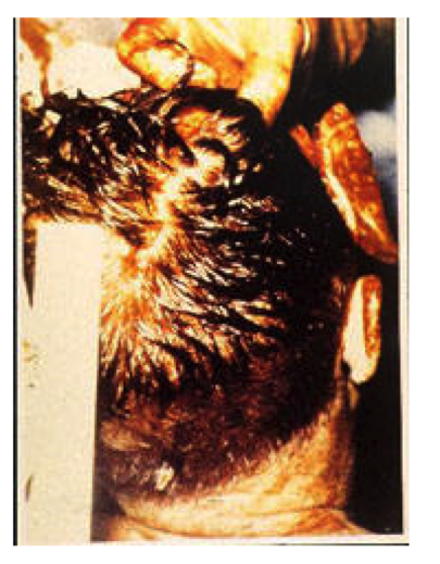
(People say this photo shows no damage, other than the small white image near the hairline, said to be adherent brain tissue. Yet just above it is an odd, light-colored, angular formation – but that is a side issue not relevant to this essay.)
What is presumed to be the wound is a flat-looking area of light, watery, reddish brown – one of many in the photo – with hairs growing out of it, apparently. If you blow it up, you will see an “X” crisscrossing through it. Possibly these are hairs.
“X” marks the spot. Hairs?
If you have ever seen an entrance wound in scalp created by a jacketed bullet traveling at medium high velocity, you will know that it looks nothing like this. The skin is crushed between bullet and bone, and appears quite dark. Lift up the scalp, and the edges of the hole are still apparent. The X’ed image in the photo does not look like a hole at all.
But this is how the wound is described on page 104 of HSCA Volume VII: “The inferior margin of this wound, from 3 to 10 o’clock, is surrounded by a crescent-shaped reddish-black area of denudation, again presenting the appearance of an abrasion collar, resulting from the rubbing of the skin by the bullet at the time of penetration. From 12 to 3 o’clock, there is a suggesting of undermining, that is, tunneling of the tissue between the skin surface and the skull…” They put this description under a drawing of the photograph.
The above description is a conflation of the drawing and photograph. I see no “reddish-black area of denudation” in the photograph – but the drawing certainly has a black and white equivalent.
Now please look at what is supposed to be an accurate drawing of this photo:
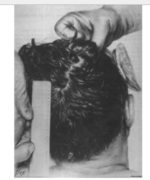
As you can see, the “wound” is much darker, has more dimension, and rolled edges. It looks like a hole at least. Not one of these characteristics appears in the photograph.
In his beautifully written essay on the medical evidence, Gary Aguilar, MD also compared the two, and said of the drawing, “the small spot visible just to the right of the top of the ruler is exaggerated in this diagram. It is significantly smaller in the original photograph.” (And there are probably other researchers who have described the contrast between the photo and the drawing.)
It is the drawing, not the photo, that John Canal presents in his articles. To the uninitiated, the drawing may appear to display a wound.
Hair Surrounding “Wound” Too Long for EOP Area
Kennedy’s hair was quite short in the back, including the area where a bullet allegedly entered. And it was even shorter below that area as it approached the neck. As you can see from the photo, the hair all around the “wound” in the cowlick is much too long.
WITNESSES DO NOT SUPPORT CANAL’S THEORY
No witnesses said pictures were taken during or after the reconstruction of the skull. None said scalp from the EOP area was dragged to the top of the head.
JOHN STRINGER
Canal’s star witness – John Stringer – who Canal insists took photographs of the reconstructed skull after the autopsy, said in 1996:
“We took no photos during or after the embalming.” (ARRB 4/8/96, p.5)
And from his longer deposition, it seems clear that Stringer only took photos during the autopsy – including the ones John Canal claims were taken after reconstruction of the skull. (ARRB 7/16/96)
But in his Washington Decoded article, Canal gives prominence to what he seems to have gotten Stringer to say in 2011, when Stringer, who was born in 1918, said at the age of 93:
“In one statement, Stringer wrote, ‘I may have taken some pictures after midnight, but I just can’t remember, it’s been too long.’ [April 30, 2011] In view of Stringer’s 1996 testimony that he did not arrive at his home (not far from the morgue) until about 4 AM, however, and that cosmetic reconstruction of the head began shortly after 11 PM, the inference that he took pictures later as well as earlier is reasonable. It is also consistent with a statement in his book MEDPHOTO that he took photos at various times throughout the procedure and whenever he was directed to do so. [21]”
Buried in Reference 21 is Stringer’s earlier statement to the ARRB:
“[21] John Stringer, MEDPHOTO: Snapshots of Life in Peace & War with the US Navy (Mooresville, NC: Wishbone Creative Product Services, 2008), 37. In an ARRB interview, Stringer also said that, ‘We took no photos during or after the embalming.’ In contrast, before that statement, he stated photos were taken “throughout the autopsy.”
Notice Canal’s last remark – as if taking pictures “throughout the autopsy” is supposed to suggest he took them after the autopsy. He pads his article with quotes from a number of people using those words, “throughout the autopsy”, as if that meant afterwards. He also conflates “late photography” to mean after the autopsy:
“Other witnesses at the postmortem whose observations support late photography [emphasis added] included Captain John Stover, an officer at the Bethesda Naval Medical Center; John Van Hoesen, one of the morticians; Joseph Hagan, supervisor of the team of morticians; Floyd Riebe, the assistant autopsy photographer; Jan Rudnicki, who assisted the autopsy doctors; General Godfrey McHugh, who observed the autopsy; Jerrol Custer, an X-ray technician; and James Sibert, one of the two FBI agents who observed the autopsy.”
Notice that he leaves out Tom Robinson, the mortician who said many things that challenged the official story.
TOM ROBINSON (mortician, not mentioned by Canal):
“When asked, Mr. Robinson said he had no recollection of photography the night of the autopsy, one way or the other – no recollection whatsoever.” (ARRB 6/21/96, p.3)
JOSEPH E. HAGAN (mortician):
“He does not recall, one way or another, whether any photographs were taken during Gawler’s work on the President’s body.” (ARRB 6/11/96 p.4)
Before the morticians worked on the body,
“Hagan said that when he arrived… the autopsy was almost over; he only had to wait in the gallery about 20 minutes before the autopsy was concluded. The body of the president was being ‘cleaned up.’ Hagen said photos were being taken, but could remember no details…” (ARRB 6/11/96, p.3)
JOHN VAN HOESEN (mortician):
Canal presents this passage from David Lifton’s book that he (Canal) says suggests photos were taken after head reconstruction. But he seems to be describing nothing more than having to wait on the autopsy and the picture-taking before they could begin their own work. And his comments seem to echo Hagen’s (see above):
“Van Hoesen: When we got up there, nothing had been started; then we had to wait for the autopsy; and then periodically, more pictures were being taken, "you know, different angles and so forth; where the entry was, and so forth; this angle, and that angle … Lifton, Best Evidence, 666.”
Canal does not report what Van Hoesen told the ARRB, and it does not help his theory any.
“He could not remember, one way or another, whether photographs were taken during the embalming and reconstruction process.” (ARRB 9/26/96, p.3)
LOGIC DOES NOT SUPPORT CANAL’S THEORY
Scalp Borrowed From an Area Missing Scalp?
It is well-established that witnesses, including a prominent brain surgeon at Parkland Hospital, said both bone and scalp were missing from an area in the back of the head that included the occiput. The lead pathologist who wrote the autopsy report, James Humes, was vague about a lot of things, including how much of the great defect involved occipital bone, but he did admit the wound was “somewhat” in that area. In any case, the back of head photograph presented earlier in this essay shows no such defect in bone or scalp.
According to Canal, scalp was borrowed from that area – even though it was already missing scalp – to cover the top right of the head.
But Canal ignores the testimony about the large hole in the scalp – which he describes as merely “torn.”
“Specifically, the cosmetic repair involved first suturing the tear in his rear scalp until it was closed, and then, after undermining, stretching the scalp until it covered the large deficit in the top/right/front of Kennedy’s head where the bullet had exited, bone was blown out, and scalp missing or badly damaged.
“The stretching of the scalp occurred after the autopsy was completed, sometime around 11 PM, and once the embalming and cosmetic restoration of the body commenced. The morticians had only the best of intentions when they took advantage of the fact that the rear scalp had only been torn, and was both repairable and useful for another purpose. They were simply trying to cover that large deficit in the head in anticipation of an open-casket funeral.” (Washington Decoded)
“The BOH opening, in all likelihood, was created after the bullet’s explosive impact exposed the president’s brain through a tear in the rear scalp and an opening between two or more dislodged (but not blown-out or missing) pieces of loose rear skull. This observation is supported by the fact that the lateral X-ray shows no missing rear bone whatsoever. Dr. J. Thornton Boswell, one of the prosectors, did say in 1996 that he repositioned some bone pieces before the X-rays and photos were taken; it seems logical that he pushed some loose pieces of skull (dislodged but still adhering to the scalp) roughly back into place. (Washington Decoded)
SUMMARY
- Not one witness mentioned by Canal says that photos were taken after the autopsy.
- In the photograph, there is no proof of a small entrance wound. But Canal does not show the photograph. He shows the drawing of it, even though the drawing, and the HSCA description of the wound, do not match the photo.
- What Canal says is the wound imported from just above the EOP is surrounded by hair that is much too long for that area.
- How Canal avoids the problem that scalp would not likely be borrowed from an area that has a sizable hole in it: he claims falsely that in the back of the head, the scalp was merely torn.
Despite the absence of proof in any of his articles, he said “The evidence for these BOH photographs being taken after the autopsy is irrefutable and so extensive it would not be practical to list here.” The SOP 2015.
What is “extensive” is the list of problems in Canal’s essays, but I focused only on those that seem to be the worst.
I wrote this essay in response to an email I received from a student at a college in Texas. She was having trouble making sense of these articles and someone referred her to me.
ADDENDUM
I have no opinion as to whether photographs were taken of a reconstructed skull, or when. I only know that witnesses do not support such a claim. While their testimony may be inaccurate or even false, it should be presented. When an author publishes a theory, the author should be the first to let the reader know whatever challenges that theory.
The testimony of Tom Robinson (excerpted below) contradicts John Canal’s assertions. Canal said the wound in the back of the head consisted only of “torn” scalp and, rather than an area of missing bone, that bone was merely displaced. He also said the bone was put back during the autopsy. But all the pathologists did was to replace loose bone that had fallen out during their probing. But they could not replace bone that was missing in the first place.
When the body was turned over to the morticians, the skull was missing a large area of bone in the back. Its appearance was not the problem; it would not have been visible during an open casket viewing. But it had to be closed to prevent embalming fluid from leaking through it.
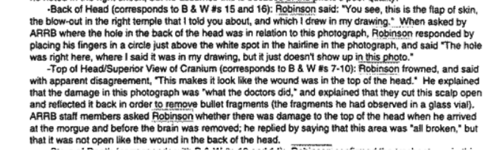
Robinson did not describe working on the top and side of the head, so we have no details about what was done in these areas – the only parts that would show in an open casket. He just said the top appeared to be “all broken” but not open like the wound in the back.
When shown the back of head photo, he said the wound was just above the white spot in the hairline. If he was right, this would mean the wound was rather low.
This is how he described the reconstruction of the area of missing bone in the back:
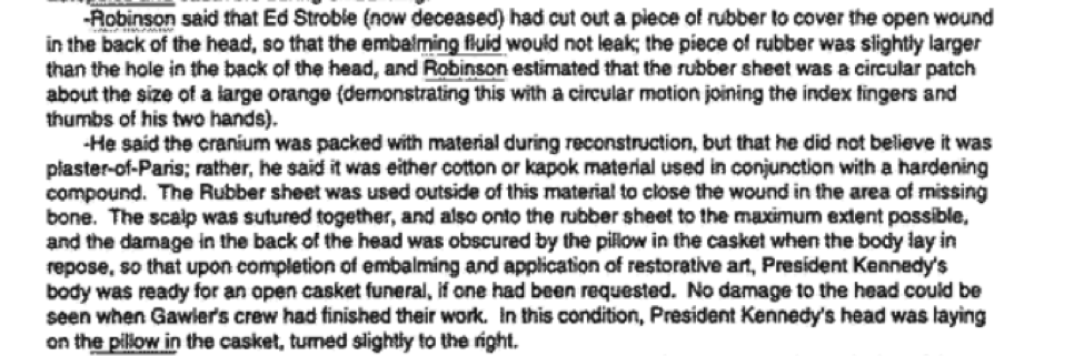
“Robinson said that Ed Stroble… had cut out a piece of rubber to cover the open wound in the back of the head… the piece of rubber was slightly larger than the hole… the rubber sheet was a circular patch about the size of a large orange… He said the cranium was packed with material during reconstruction… The rubber sheet was used outside of this material to close the wound in the area of missing bone. The scalp was sutured together, and also onto the rubber sheet to the maximum extent possible and the damage in the back of the head was obscured by the pillow in the casket…”
Location of Hole in Back; Condition of Top of Head
Size of Area of Missing Bone, Reconstruction